Smooth Muscle Diagram Labeled - Muscular System Accessscience From Mcgraw Hill Education : The skeletal muscle fibers are elongated, cylindrical and multinucleated cells whose length may vary in different animals.
byAdmin•
0
Smooth Muscle Diagram Labeled - Muscular System Accessscience From Mcgraw Hill Education : The skeletal muscle fibers are elongated, cylindrical and multinucleated cells whose length may vary in different animals.. In this video i have shown the simplest way of drawing muscle drawing. Diagram of smooth muscle cardiac muscle and straited muscle tissue. In skeletal muscle, a single type of somatic nervous system traverses to muscle, where it stimulates organelle in the muscle cells in order to release calcium. Individual muscle fibers, (b) surrounds groups of skeletal muscle fibers (fascicles), and (c) covers the muscle as a whole. Muscle anatomy coloring sheets 12 photos of the muscle anatomy coloring sheets free muscle anatomy coloring sheets, muscle anatomy coloring pages, muscle anatomy coloring pages free, muscle anatomy coloring sheets, human muscles, free muscle anatomy coloring sheets, muscle anatomy coloring pages, muscle anatomy.
It constitutes much of the musculature of Smooth muscles have a much stronger ability to contract than skeletal. The muscles of the human body can be categorized into a number of groups which include muscles relating to the head and neck, muscles of the torso or trunk, muscles of the upper limbs, and muscles of the lower limbs. Skeletal muscle tissue is composed of long cells called muscle fibers that have a striated appearance. Myocardial to smooth muscle junction of the arterial pole whole.
Histology 4000 Muscle Lecture Notes 6 from www.auburn.edu Smooth muscle cells are found in the dividers of empty organs, including the stomach, digestion tracts, urinary bladder and uterus, and in the dividers of paths, for example, the supply routes and veins of the circulatory framework, and the tracts of the. Kierszenbaum, al histology and cell biology 2nd ed., mosby elsevier, 2007, p. Name the tough connective tissue cord that serves to attach a muscle to a bone. Skeletal muscle tissue is composed of long cells called muscle fibers that have a striated appearance. Draw a labeled diagram of smooth muscle. Myocardial to smooth muscle junction of the arterial pole whole. They range from about 30 to 200 μm (thousands of times shorter than skeletal muscle fibers), and they produce their own connective tissue, endomysium.although they do not have striations and sarcomeres, smooth muscle fibers do have actin and myosin. Smooth muscle tissue, unlike striated muscle, contracts slowly and automatically.
Structure and composition of muscle meat science.
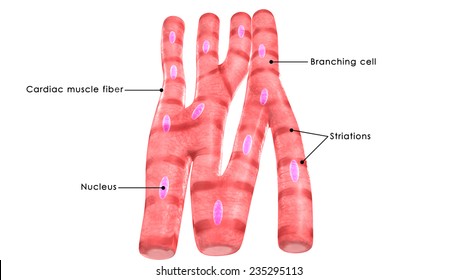
In this short guide, you will get a basic concept of skeletal muscle histology from the real slide and labeled diagram. Name the tough connective tissue cord that serves to attach a muscle to a bone. Schematic diagram of cardiac muscle click to see enlarged view: Individual muscle fibers, (b) surrounds groups of skeletal muscle fibers (fascicles), and (c) covers the muscle as a whole. 1 response to simple smooth muscle diagram labeled unknown june 5, 2021 at 12:39 am. Myocardial to smooth muscle junction of the arterial pole whole. Smooth muscle cells are found in the dividers of empty organs, including the stomach, digestion tracts, urinary bladder and uterus, and in the dividers of paths, for example, the supply routes and veins of the circulatory framework, and the tracts of the. Draw a labeled diagram of smooth muscle. Skeletal muscles attach to and move bones by contracting and relaxing in response to voluntary messages from the nervous system. It constitutes much of the musculature of Smooth muscle, muscle that shows no cross stripes under microscopic magnification. Intercalated disk (ventricle, cat) click to see enlarged view: Each slide has descriptions and images with a task for students to perform, such as labeling a diagram (based on the description) or.
A gap junction (gj) is present in the longitudinal portion of the disk. The skeletal muscle fibers are elongated, cylindrical and multinucleated cells whose length may vary in different animals. Draw a labeled diagram of smooth muscle. Muscle fibers are organized into bundles supplied by blood vessels and innervated by motor neurons. Name three types of fiber arrangements seen in skeletal muscle.
Novel Myh11 Dual Reporter Mouse Model Provides Definitive Labeling And Identification Of Smooth Muscle Cells Brief Report Arteriosclerosis Thrombosis And Vascular Biology from www.ahajournals.org Related posts of smooth muscle labelled diagram muscle anatomy shoulder. A gap junction (gj) is present in the longitudinal portion of the disk. Schematic diagram of cardiac muscle click to see enlarged view: In this video i have shown the simplest way of drawing muscle drawing. Smooth muscle cells are found in the dividers of empty organs, including the stomach, digestion tracts, urinary bladder and uterus, and in the dividers of paths, for example, the supply routes and veins of the circulatory framework, and the tracts of the. They range from about 30 to 200 μm (thousands of times shorter than skeletal muscle fibers), and they produce their own connective tissue, endomysium.although they do not have striations and sarcomeres, smooth muscle fibers do have actin and myosin. It constitutes much of the musculature of Muscle anatomy shoulder 12 photos of the muscle anatomy shoulder muscle anatomy of the shoulder and neck, muscle anatomy shoulder upper arm, shoulder muscle anatomy game, shoulder muscle anatomy posterior view, shoulder muscle anatomy workout, human muscles, muscle anatomy of the shoulder and neck, muscle anatomy.
1 response to simple smooth muscle diagram labeled unknown june 5, 2021 at 12:39 am.
Draw a labeled diagram of smooth muscle. Myocardial to smooth muscle junction of the arterial pole whole. You will also get the identification points of skeletal muscle histology slide with a little description here in this guide. Muscle anatomy shoulder 12 photos of the muscle anatomy shoulder muscle anatomy of the shoulder and neck, muscle anatomy shoulder upper arm, shoulder muscle anatomy game, shoulder muscle anatomy posterior view, shoulder muscle anatomy workout, human muscles, muscle anatomy of the shoulder and neck, muscle anatomy. Name three types of fiber arrangements seen in skeletal muscle. Kierszenbaum, al histology and cell biology 2nd ed., mosby elsevier, 2007, p. Skeletal muscles attach to and move bones by contracting and relaxing in response to voluntary messages from the nervous system. Muscle anatomy coloring sheets 12 photos of the muscle anatomy coloring sheets free muscle anatomy coloring sheets, muscle anatomy coloring pages, muscle anatomy coloring pages free, muscle anatomy coloring sheets, human muscles, free muscle anatomy coloring sheets, muscle anatomy coloring pages, muscle anatomy. Structure and composition of muscle meat science. Schematic diagram of cardiac muscle click to see enlarged view: Smooth muscle, muscle that shows no cross stripes under microscopic magnification. The diagram is fully labeled. This activity was created for distance learning during the 2020 pandemic as a substitution for traditional dissections and lessons that involve identifying the muscles and their function.
Structure and composition of muscle meat science. 12 photos of the smooth muscle diagram. Smooth muscle (textus muscularis levis) smooth muscle is a type of tissue found in the walls of hollow organs, such as the intestines, uterus and stomach. Schematic diagram of cardiac muscle click to see enlarged view: Smooth muscle cells are found in the dividers of empty organs, including the stomach, digestion tracts, urinary bladder and uterus, and in the dividers of paths, for example, the supply routes and veins of the circulatory framework, and the tracts of the.
Smooth Muscle Hd Stock Images Shutterstock from image.shutterstock.com Related posts of smooth muscle diagram labeled muscle anatomy coloring sheets. The diagram is fully labeled. Skeletal muscles attach to and move bones by contracting and relaxing in response to voluntary messages from the nervous system. Individual muscle fibers, (b) surrounds groups of skeletal muscle fibers (fascicles), and (c) covers the muscle as a whole. 12 photos of the smooth muscle diagram. Skeletal muscles, smooth muscles, cardiac muscles. It is the pen diagram of skeletal, smooth and cardiac muscle for class 10, 11 and 12. The muscles of the human body can be categorized into a number of groups which include muscles relating to the head and neck, muscles of the torso or trunk, muscles of the upper limbs, and muscles of the lower limbs.
In skeletal muscle, a single type of somatic nervous system traverses to muscle, where it stimulates organelle in the muscle cells in order to release calcium.
Intercalated disk (ventricle, cat) click to see enlarged view: Schematic diagram of cardiac muscle click to see enlarged view: Related posts of smooth muscle diagram labeled muscle anatomy coloring sheets. Skeletal muscles attach to and move bones by contracting and relaxing in response to voluntary messages from the nervous system. Smooth muscle tissue, unlike striated muscle, contracts slowly and automatically. Diagram of smooth muscle contraction, smooth cardiac and skeletal muscle diagram, smooth muscle cell diagram, smooth muscle cell picture, smooth muscle contraction diagram, human muscles, diagram of smooth muscle contraction, smooth cardiac and skeletal muscle diagram, smooth muscle cell diagram, smooth. Smooth muscles are unique in their largely involuntary response, and in their structure. Skeletal muscles, smooth muscles, cardiac muscles. Smooth muscles are unique in their largely involuntary response, and in their structure. Smooth muscles have a much stronger ability to contract than skeletal. This activity was created for distance learning during the 2020 pandemic as a substitution for traditional dissections and lessons that involve identifying the muscles and their function. Structure and composition of muscle meat science. Smooth muscle makes up the walls of hollow organs, respiratory passageways, and blood vessels.